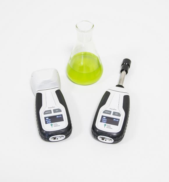
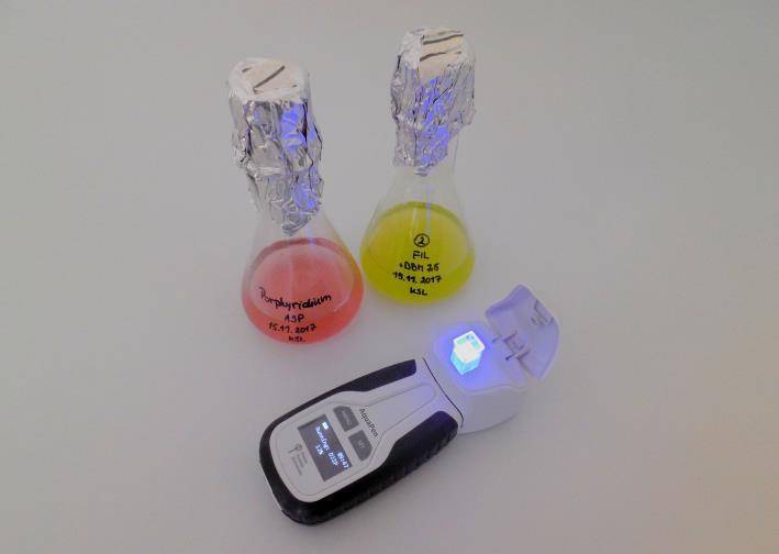
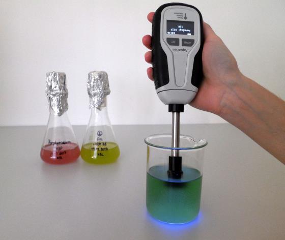
AquaPen AP110手持式藻类荧光测量仪是一款用于快速、精确测量水体藻类与蓝藻叶绿素荧光参数的手持式荧光仪。AquaPen有两种探头型号。AP110-C配备比色杯试管测量室,将要测量的水体、悬浊液或培养溶液采集到比色杯中进行测量,配备455nm蓝色和620nmLED红色光源,既可以测量叶绿素荧光,又可以测量680nm和720nm光密度。AP110-P配备了浸入式光学探头,可直接插到要测量的水体、悬浊液或培养溶液中进行测量,也可测量大型藻类。
AquaPen 具备极高的敏感度,可检测0.5μg Chl/L的叶绿素荧光,可以检测浮游植物浓度极低的自然水体,可用于野外和实验室测量。
AquaPen采用调试式荧光测量技术,可设置多种参数,方便测量多种植物叶绿素荧光。外观小巧,方便携带,设计新颖,操作简单,经济耐用,精度高稳定性好。
应用领域
藻类、蓝藻光合特性研究
水体藻类含量检测
光合突变体筛选与表型研究
生物和非生物胁迫的检测
藻类抗胁迫能力或者易感性研究
经济藻类育种、病害检测、长势与产量评估
教学
功能特点:
结构紧凑、便携性强,LED光源、检测器、控制单元集成于仅手机大小的仪器内,重量仅180g
功能强大,是叶绿素荧光技术的高端结晶产品,具备了大型荧光仪的所有功能,可以测量所有叶绿素 荧光参数
内置了所有通用叶绿素荧光分析实验程序,包括两套荧光淬灭分析程序、3套光响应曲线程序、OJIP–test等
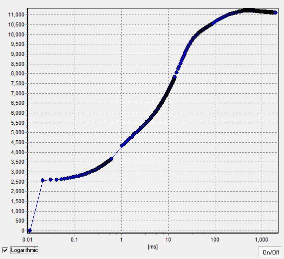
高时间分辨率,可达10万次每秒,自动绘出OJIP曲线并给出26个OJIP–test参数
AquaPen两种探头型号:比色杯试管测量室,既可以测量叶绿素荧光,又可以测量680nm和720nm光密度;浸入式光学探头,可直接插到要测量的水体、悬浊液或培养溶液中进行测量,也可测量大型藻类
FluorPen专业软件功能强大,可下载、展示叶绿素荧光参数图表,也可以通过软件直接控制仪器进行测量
具备无人值守自动监测功能
内置蓝牙与USB双通讯模块, GPS模块,输出带时间戳和地理位置的叶绿素荧光参数图表
配备多种叶夹型号:固定叶夹式(适用于大批量样品快速测量)、分离叶夹式(适用于暗适应测量)、开放叶夹式(适用于温室、培养箱进行监测)、用户定制式等
可选配野外自动监测式荧光仪,防水防尘设计
测量程序与功能
Ft:瞬时叶绿素荧光,暗适应完成后Ft=F0
QY:量子产额,表示光系统II 的效率,等于Fv/Fm(暗适应状态)或ΦPSII (光适应状态)。
OJIP:快速荧光动力学曲线,用于研究植物暗适应后的快速荧光动态变化
NPQ:荧光淬灭动力学曲线,用于研究植物从暗适应到光适应状态的荧光淬灭变化过程。
LC:光响应曲线,用于研究植物对不同光强的荧光淬灭反应。
OD:光密度,反映藻类密度(限AP110-C)。
技术参数
测量参数包括F0、Ft、Fm、Fm’、QY、QY_Ln、QY_Dn、NPQ、Qp、Rfd、Area、Mo、Sm、PI、ABS/RC等50多个叶绿素荧光参数,OD680和OD720(限AP110-C)及3种给光程序的光响应曲线、2种荧光淬灭曲线、OJIP曲线等
OJIP–test时间分辨率为10µs(每秒10万次),给出OJIP曲线和26个参数,包括F0、Fj、Fi、Fm、Fv、Vj、Vi、Fm/F0、Fv/F0、Fv/Fm、Mo、Area、Fix Area、Sm、Ss、N、Phi_Po、Psi_o、Phi_Eo、Phi–Do、Phi_Pav、PI_Abs、ABS/RC、TRo/RC、ETo/RC、DIo/RC等
测量程序:Ft、QY、OJIP、NPQ1、NPQ2、LC1、LC2、LC3、OD(限AP110-C)、Multi无人值守自动监测
测量光:每测量脉冲0-0.09µmol(photons)/m².s,0-可调
光化学光:0–1000µmol(photons)/m².s,0-可调
饱和光:0–3000µmol(photons)/m².s,0-可调
探头型号:AP110-C试管式、AP110-P探头式

光源:AP110-C:620nm红光和455nm蓝光测量叶绿素荧光,680nm和720nm红外光测量OD;AP110-P:455nm蓝光
试管容积(限AP110-C):4ml
叶绿素荧光检测限:0.5μg Chl/L
检测器:PIN光电二极管,667–750nm滤波器
尺寸大小:超便携,手机大小,165×65×55mm,重量仅290g
存贮:容量16Mb,可存储149000数据点
显示与操作:图形化显示,双键操作,待机8分钟自动关闭
供电:可充电锂电池,USB充电,连续工作48小时,低电报警
工作条件:0–55℃,0–95%相对湿度(无凝结水)
存贮条件:-10–60℃,0–95%相对湿度(无凝结水)
通讯方式:蓝牙 USB双通讯模式
GPS模块:内置
软件:FluorPen1.1专用软件,用于数据下载、分析和图表显示,输出Excel数据文件及荧光动力学曲线图,适用于Windows 7及更高操作系统
操作软件与实验结果

产地:
欧洲
参考文献
1. M Zhao, et al. 2019. Effects of dissolved organic matter from different sources on Microcystis aeruginosa growth and physiological characteristics. Ecotoxicology and Environmental Safety 176: 125-131
2. L Yang, et al. 2019. Biotoxicity of water-soluble species in PM2.5 using Chlorella. Environmental Pollution 250: 914-921
3. Y Li, et al. 2019. Darkness and low nighttime temperature modulate the growth and photosynthetic performance of Ulva prolifera under lower salinity. Marine Pollution Bulletin 146: 85-91
4. P Ranadheer, et al. 2019. Non-lethal nitrate supplementation enhances photosystem II efficiency in mixotrophic microalgae towards the synthesis of proteins and lipids. Bioresource Technology 283: 373-377
5. M Jiang, et al. 2019. Allelopathic effects of harmful algal extracts and exudates on biofilms on leaves of Vallisneria natans. Science of The Total Environment 655: 823-830
6. PV Sijil, et al. 2019. Enhanced accumulation of alpha-linolenic acid rich lipids in indigenous freshwater microalga Desmodesmus sp.: The effect of low-temperature on nutrient replete, UV treated and nutrient stressed cultures. Bioresource Technology 273: 404-415
7. W Noh, et al. 2019. The potential of a natural biopolymeric flocculant, ε-poly-l-lysine, for harvesting Chlorella ellipsoidea and its sustainability perspectives for cost and toxicity. Bioprocess and Biosystems Engineering 42(6): 971-978
8. M Bao, et al. 2019. Different Photosynthetic Responses of Pyropia yezoensis to Ultraviolet Radiation Under Changing Temperature and Photosynthetic Active Radiation Regimes. Photochemistry and Photobiology, doi: 10.1111/php.13108
9. G Konert, et al. 2019. Protein arrangement factor: a new photosynthetic parameter characterizing the organization of thylakoid membrane proteins. Physiologia Plantarum 166: 264-277.
10. Z Zhu, et al. 2019. High copper and UVR synergistically reduce the photochemical activity in the marine diatom Skeletonema costatum. Journal of Photochemistry and Photobiology B: Biology 192: 97-102
11. X Chen, et al. 2018. The secretion of organics by living Microcystis under the dark/anoxic condition and its enhancing effect on nitrate removal. Chemosphere 196: 280-287
12. C M'Rabet, et al. 2018. Impact of two plastic-derived chemicals, the Bisphenol A and the di-2-ethylhexyl phthalate, exposure on the marine toxic dinoflagellate Alexandrium pacificum. Marine Pollution Bulletin 126: 241-249
13. P Steinrücken, et al. 2018. Comparing EPA production and fatty acid profiles of three Phaeodactylum tricornutum strains under western Norwegian climate conditions. Algal Research 30: 11-22
14. T Kieselbach, et al. 2018. Proteomic analysis of the phycobiliprotein antenna of the cryptophyte alga Guillardia theta cultured under different light intensities. Photosynthesis Research 135(1-3): 149-163
15. SB Ouada, et al. 2018. Effect and removal of bisphenol A by two extremophilic microalgal strains (Chlorophyta). Journal of Applied Phycology 6: 1-12